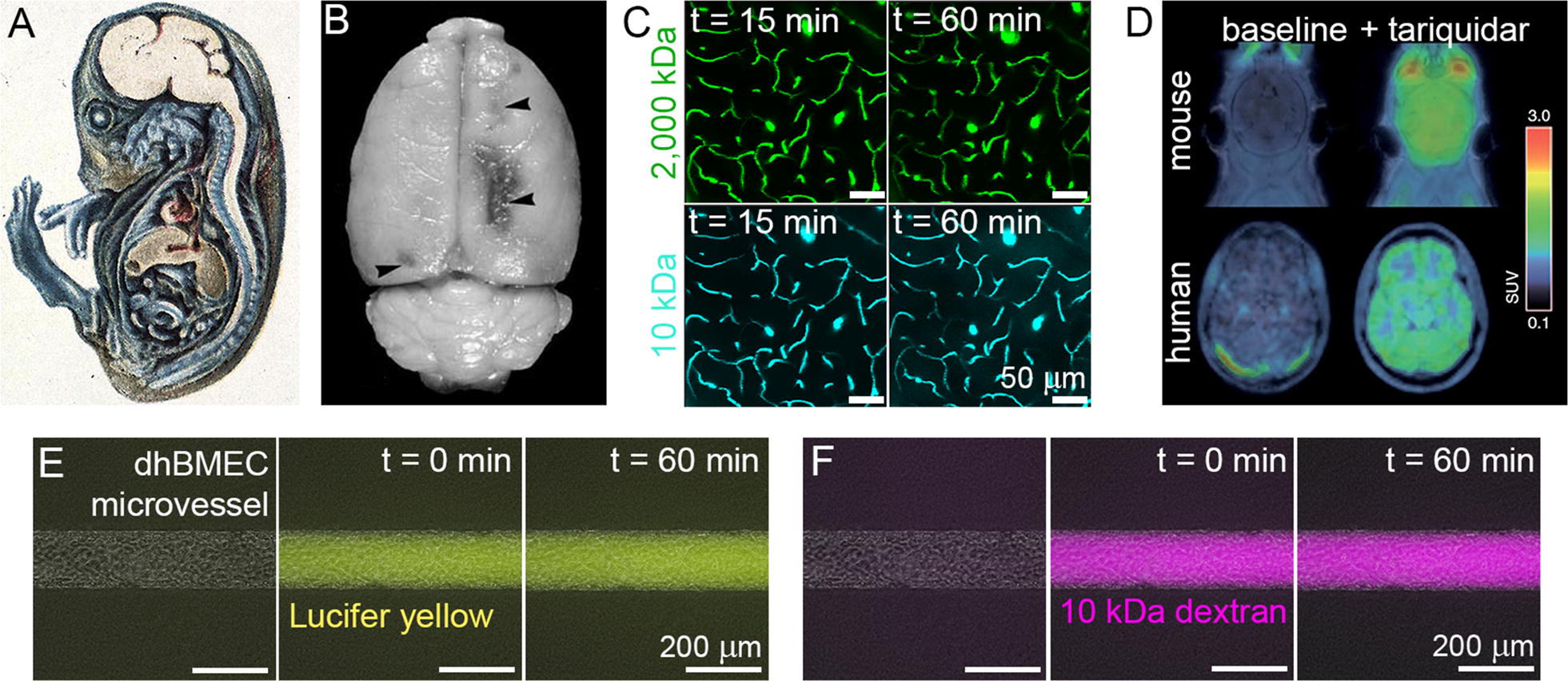
Fig. 4.

Blood–brain barrier permeability. A Guinea pig embryo injected with trypan blue demonstrates restriction of dye entry into CNS (from [135]). B Brain of a rat with chronic hypertension showing areas of Evans blue extravasation in the boundary zone areas (from [141]). C Adult mice injected with fluorescently-labeled dextrans (10 and 2000 kDa) and imaged with two-photon microscopy show lack of significant dye extravasation over 1 h (from [142]). D Positron emission tomography (PET) imaging of radiolabeled verapamil in mouse and human brains with and without p-glycoprotein inhibition using tariquidar. The color bar indicates brain-to-plasma ratio, a measure of drug penetration (from [143]). E, F Permeability experiments using Lucifer yellow and 10 kDa dextran in tissue-engineered iPSC-derived brain microvessels (from [24])
