Fig. 1.
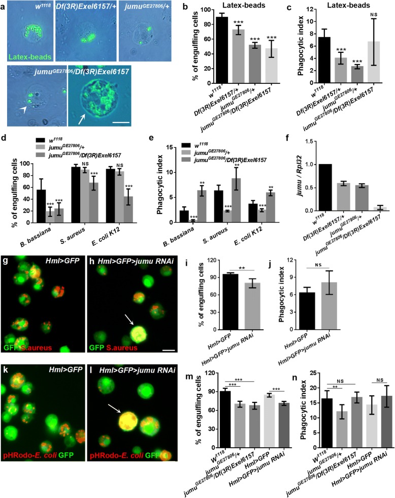
Loss of jumu results in defective phagocytosis in circulating hemocyte. a, g, h, k, l Circulating hemocytes were isolated from third-instar larvae 1 h after injection with latex beads, B. bassiana, E. coli, S. aureusat or pHRodo-E. coli. b, d, i, m Quantification of the percentage of engulfing cells in the phagocytosis assays of circulating hemocytes. c, e, j, n Quantification of phagocytosis indexes (the number of engulfed latex beads or bacteria per hemocyte) in phagocytosis assays of circulating hemocytes. f Real-time PCR analysis of the jumu level in the entire third-instar larvae. For all quantifications, the error bars represent ±S.E.M of at least 3 independent experiments; NS, not significant; **P < 0.01; ***P < 0.001 (Student’s t-test). The arrowhead in a indicates the jumuGE27806/Df(3R)Exel6157 circulating hemocytes with normal size. The arrows in a, g and k indicate the enlarged circulating hemocytes resulting from the loss of jumu. Scale bars: 10 μm

