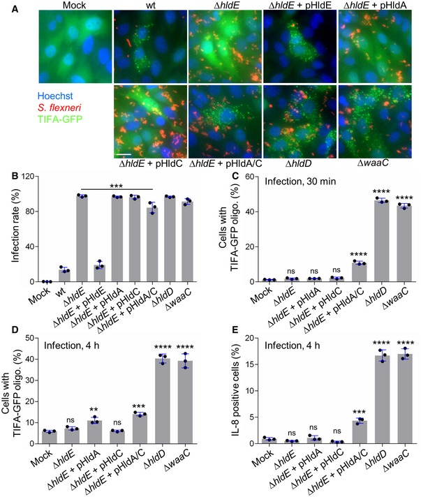
Representative images of the formation of TIFA oligomers in HeLa cells infected for 30 min with dsRed‐expressing wt or indicated mutants of S. flexneri (MOI 50). pHldA/C means plasmid‐encoded HldA and HldC. Fluorescence intensity was adjusted between strains to optimize visualization of bacteria. Scale bar, 20 μm.
Infectivity of indicated S. flexneri strains.
TIFA‐GFP oligomerization after 30 min of infection with selected mutants of the LPS biosynthesis pathway (MOI 10).
TIFA‐GFP oligomerization after 4 h of infection with the S. flexneri mutants shown in (C).
IL‐8 production 4 h postinfection (MOI 5).
Data information: In (B–E), data correspond to the mean ± SD of three independent experiments. For comparison between mock and each infected condition (C–E) or Δ
hldE and double‐complemented mutant (B), statistical significance was assessed using one‐way ANOVA followed by Tukey's multiple comparisons test **
P < 0.01, ***
P < 0.001, ****
P < 0.0001, not significant (ns).

