
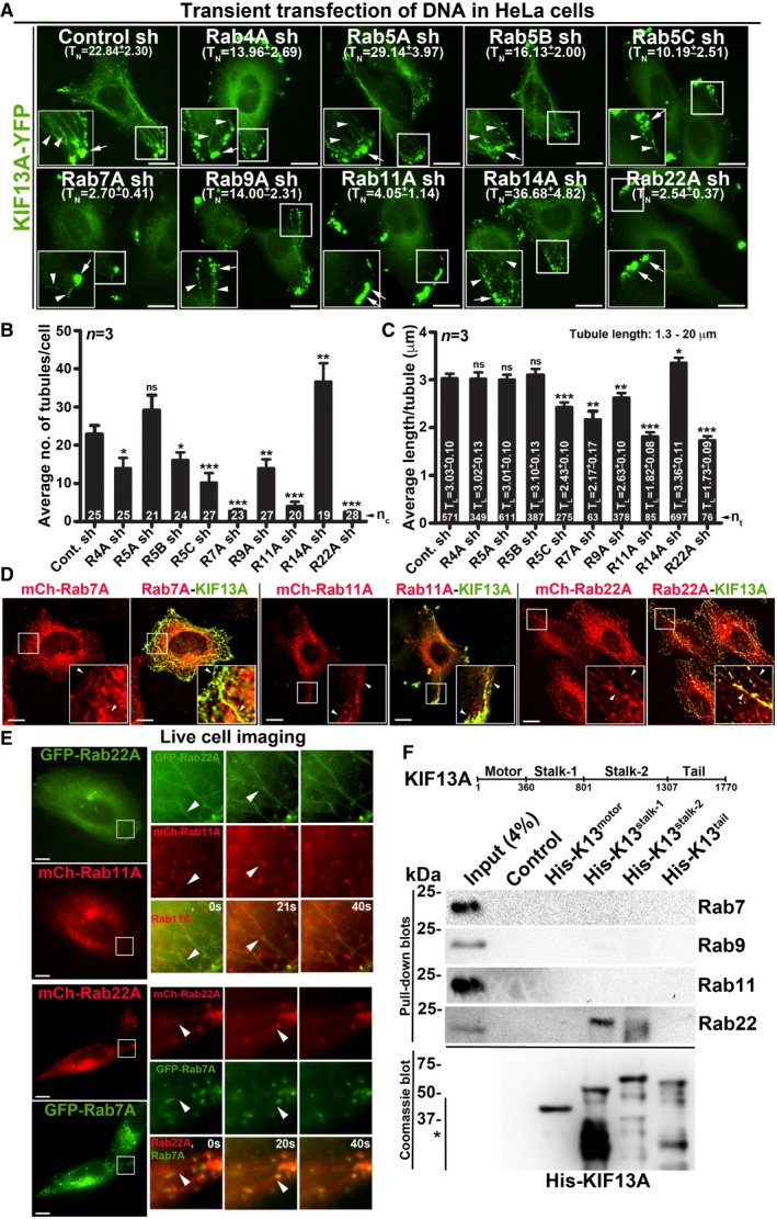
Figure 1. Selected endosomal Rab RNAi screen identified Rab22A as a regulator of RE dynamics.
-
AIFM images of KIF13A‐YFP‐transfected control and Rab‐knockdown HeLa cells. TN: average tubule number (mean ± SEM, n = 3).
-
B, CGraphs represent the measurement of KIF13A‐positive TN (B) and TL (C) in HeLa cells of Fig 1A (mean ± SEM). n = 3. nc: total number of cells. nt: total number of tubules. *P ≤ 0.05, **P ≤ 0.01, ***P ≤ 0.001 and ns = not significant (unpaired Student's t‐test).
-
DIFM images of KIF13A‐YFP and mCherry‐Rab7A/11A/22A‐transfected HeLa cells.
-
ELive cell imaging of GFP/mCherry‐Rab22A with respect to mCherry‐Rab11A or GFP‐Rab7A in HeLa cells. Magnified view of insets (at 0, 20, 40 s) are shown separately.
-
FPull‐down of different His‐KIF13A domains using HeLa cell lysate and then probed with indicated Rab proteins. The bead‐bound His‐KIF13A in each pull‐down was shown on the Coomassie‐stained gel. *, non‐specific bands. Note, part of this experiment was shown in Fig 5F.