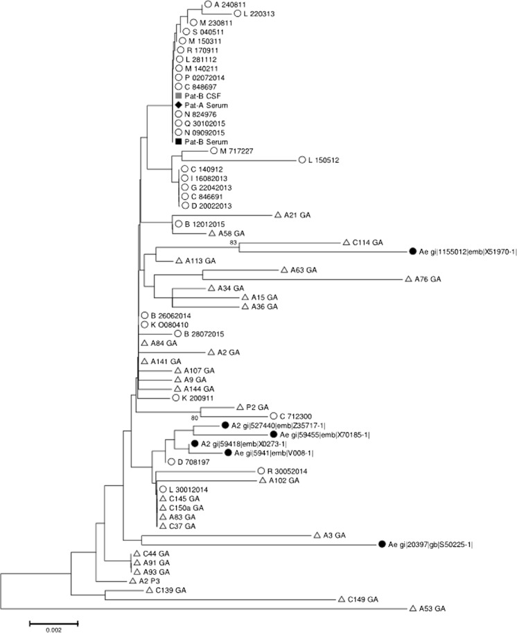
Fig. 1.
Phylogenetic tree of 721 nucleotide polymerase coding HBV genotype A sequences (n = 67). Open circle: not related randomly selected local sequences (n = 29); open triangle: acute French hepatitis B (n = 29); closed circle: Genbank reference sequences (n = 6); closed losange: patient A blood strain; closed square and gray square: patient B blood and CSF strains, respectively. The evolutionary distances were computed using the Jukes-Cantor method. The percentage of replicate trees in which the associated taxa clustered together in the bootstrap test (500 replicates) is shown next to the branches when above 70%

