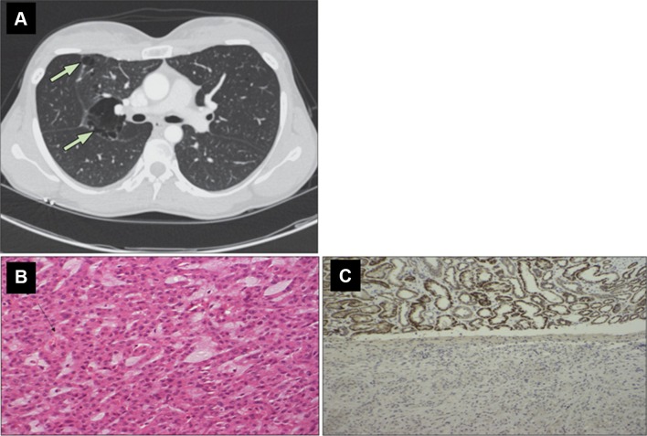
Fig. 1.
Examples of radiological, histological and immunohistochemical features that might suggest an inherited predisposition to renal cell carcinoma. Upper panel: a high-resolution CT thorax showing multiple basal cysts in a patient with Birt–Hogg–Dube syndrome (reprinted with permission from [52]). Lower panel: b the H + E-stained histological appearance of an SDHB-deficient RCC. There is evidence of intracytoplasmic vacuoles marked by the black arrow. c Loss of SDHB protein expression on immunostaining of the RCC tumour in the lower part of the image, with SDHB staining present in the adjacent normal renal tissue visible in the upper image.
(Reprinted with permission from [39])

