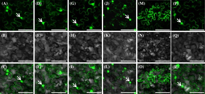
Fig. 3.
Subcellular localization of Physalis EJC core proteins in plant cells. a–c PFMAGO1-GFP. d–f PFMAGO2-GFP. g–i PFY14-GFP. j–l PFeIF4AIII-GFP. m–o PFBTZ-GFP. p–r GFP protein as the control. Bars, 50 µm. The first row shows signals in fluorescence fields; the second row shows signals in bright fields; the third row shows merged signals of fluorescence and bright fields. The arrow indicates the nucleus

