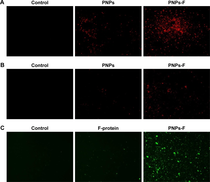
Figure 2.
In vitro uptake of PNPs-F by immune cells.
Notes: (A) PBMCs and (B) splenocytes were treated with medium or RITC dye-tagged PNPs, or RITC dye-tagged PNPs-F for 4 hours and examined in red channel under fluorescent microscopy at 20× magnification. (C) PBMCs were treated with medium or F-protein, or PNPs-F for 4 hours and immunostained with flagellin antibody, and cells were observed in green channel under fluorescent microscopy at 20× magnification.
Abbreviations: F, flagellar; PBMCs, peripheral blood mononuclear cells; PNPs, polyanhydride nanoparticles; RITC, rhodamine B isothiocyanate; PNPs-F, surface F-protein-coated PNPs.

