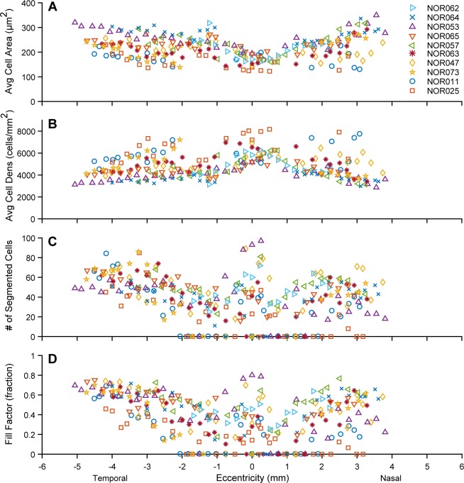
Figure 3.
Measured SWAF RPE cell statistics for participants, listed in order of increasing age in the legend. Cell area increases with eccentricity (A), where the rate of increase differed between participants. Measured cell densities (B) in the three oldest participants were among the highest, though these cells were typically difficult to segment. (C, D) Cell segmentation was consistently greater in the periphery, particularly temporal locations, while the fovea exhibited high variability. Eighty-nine percent of ROIs with no segmented cells fell within ± 2 mm of the fovea and in the nasal retina, perhaps because of increased absorption and scatter due to melanin, vasculature, and/or maculopapillary nerve fiber bundles (in the nasal retina).

