Figure 8.
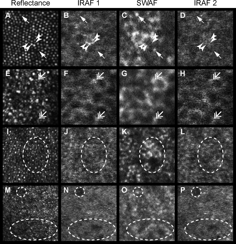
Microscopic SWAF and IRAF structure at the fovea (A–D) and 10° temporally (E–H) for participant NOR053, 10° temporally (I–L) in NOR063, and near foveal center (M–P) of NOR025. From left to right, columns show the NIR reflectance image, the first acquired IRAF image (IRAF 1), the SWAF image, and the second acquired IRAF image (IRAF 2). Incidences of colocalized (double head arrows) and noncolocalized (arrows, arrowheads) punctate hyper-AF between modalities were observed. Four distinct points of hyper-IRAF (arrowheads) in (B) have the same size and location of cones in reflectance (A), and are absent in the IRAF 2 image (D) acquired 1.3 hours later. The dashed oval in (K) shows a SWAF patch of hyper-AF and hypo-AF, while the IRAF images show no corresponding features at the same location. In contrast, (O) shows two patches (dashed circle and oval) of hyper-SWAF that appear to be hypo-AF in IRAF. (A–H) 75 × 75 μm. (I–P) 150 × 150 μm.

