Figure 5.
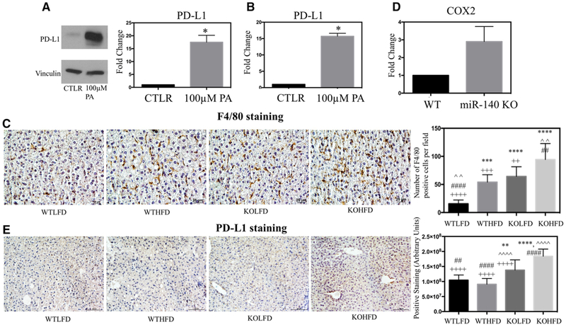
High-fat diet and miR-140 loss promote an inflammatory hepatic microenvironment. A) Western blotting demonstrating protein expression of PD-L1 in primary human hepatocytes following treatment with 100 um palmitic acid for 24 h. B) qRT-PCR of PD-L1 expression in primary hepatocytes following treatment with 100 um palmitic acid for 24 h. C) Immunohistochemistry staining for pan-macrophage marker F4/80. Quantification performed in ImageJ, average number of macrophage Kupffer cells per field. p < 0.0001. D) qPCR detection of COX-2 expression in primary peritoneal macrophages isolated from wild-type and miR-140 knockout mice. E) Immunohistochemistry staining for PD-L1. Quantification performed in ImageJ. p < 0.0001. *, comparison with WTLFD. ˆ, comparison with WTHFD. #, comparison with KOLFD.+, comparison with KOHFD. p-Value is indicated as: *p < 0.05. **p < 0.01. ***p < 0.001. ****p < 0.0001.

