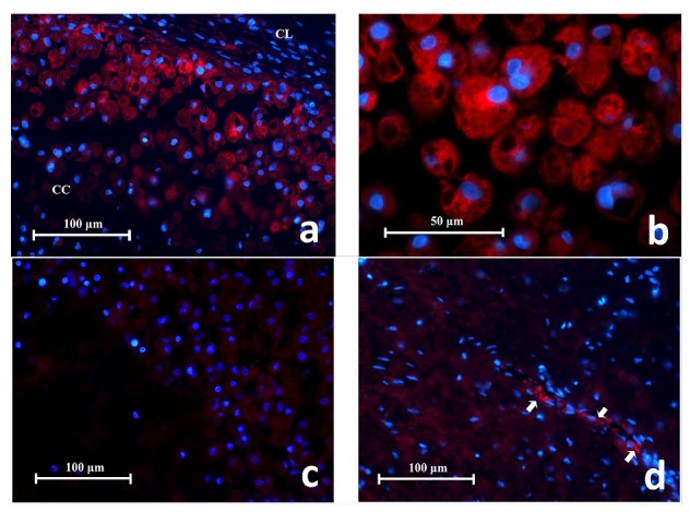
Figure 7.

( a) Representative histopathology section showing CD68 + cells in a mucocele fluorescing in red. They are found in large number adjacent to the cystic lining (CC) and spread into the cystic cavity (CC). Characteristics of macrophage cells are evident e.g. irregular cell shape, intra-cellular vacuoles (x400). ( b) High power magnification of CD68 + cells in a mucocele showing their large spongy globular appearance. The red fluorescence is observed on the cell membrane and cytoplasm but not the intracellular vesicles or phagosomes (x1000). ( c) Representative section of negative antibody control. ( d) Representative section of negative tissue control (periapical scar) with autofluorescing RBCs (white arrows) (x400).
