Figure 3.
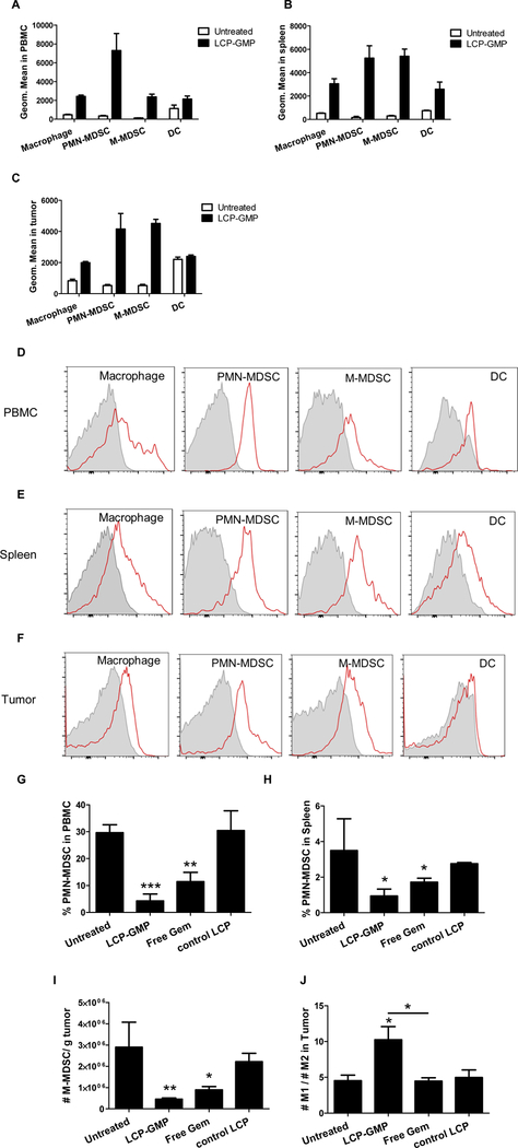
LCP-GMP was effectively taken up by myeloid cells and significantly eliminated MDSCs in the peripheral blood, spleen and tumor, as well as induced macrophage polarization towards anti-tumor M1 phenotype in tumors. (A-C) The myeloid cell uptake of LCP-GMP in the (A) peripheral blood, (B) spleen and (C) tumor was analyzed at 12 h after i.v. injection of NBD-labeled LCP-GMP into B16F10 tumor-bearing mice. Leukocytes in the peripheral blood, spleen and tumor were stained with antibodies against different myeloid cell populations (macrophage, PMN-MDSC, M-MDSC, DC). Cellular uptake of NBD-labeled LCP-GMP in myeloid cells was analyzed by flow cytometry. The representative FACS histograms of cellular uptake of LCP-GMP in different myeloid cell populations in the (D) peripheral blood, (E) spleen and (F) tumor at 12 h post injection were shown. Gray: Untreated. Red: LCP-GMP. B16F10 tumor-bearing mice were given i.v. injections of LCP-GMP, free Gem and control LCP on days 8, 10, 12, 14 post tumor cell inoculation. On day 16, MDSCs in peripheral blood, spleens and tumors were analyzed by flow cytometry. (G) The frequencies of PMN-MDSCs in peripheral blood after treatments. **p<0.01, Untreated vs. Free Gem; ***p<0.0005, Untreated vs. LCP-GMP. (H) The frequencies of PMN-MDSCs in spleens after treatments. *p<0.05, Untreated vs. LCP-GMP and Untreated vs. Free Gem. (I) The numbers of M-MDSCs in tumors after treatments. Data were normalized to tumor weights. *p<0.05, Untreated vs. Free Gem; **p<0.005, Untreated vs. LCP-GMP. (J) The ratios of anti-tumor M1 to pro-tumor M2 macrophages in tumors after treatments. *p<0.05, Untreated vs. LCP-GMP and LCP-GMP vs. Free Gem. (n=4 per group)

