
Figure 4.
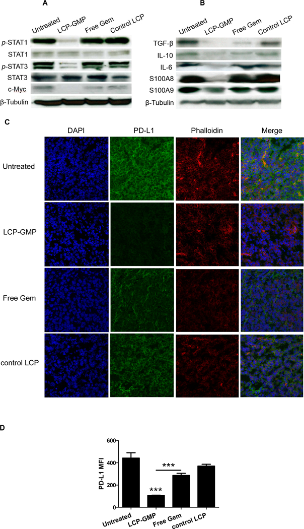
LCP-GMP downregulated the expressions of pro-tumor (A) transcription factors and (B) immunosuppressive mediators as well as (C-D) PD-L1 in tumors after treatments. B16F10 tumor-bearing mice were given i.v. injections of LCP-GMP, free Gem and control LCP on days 8, 10, 12, 14 post tumor cell inoculation. On day 16, tumor lysates were prepared for western blot analysis (A-B), and frozen sections from dissected tumors were prepared for immunostaining (C-D). (C) The PD-L1 expressions in tumors after treatments were detected by immunohistochemistry before observing under a confocal microscope (n=4 per group). Blue: nuclei; green: PD-L1; red: phalloidin. (D) The mean fluorescence intensities (MFI) of PD-L1 signals in confocal images were analyzed by ImageJ software, as indicators of the expression levels of PD-L1 in tumors after treatments. ***p<0.0001, Untreated vs. LCP-GMP and LCP-GMP vs. Free Gem.