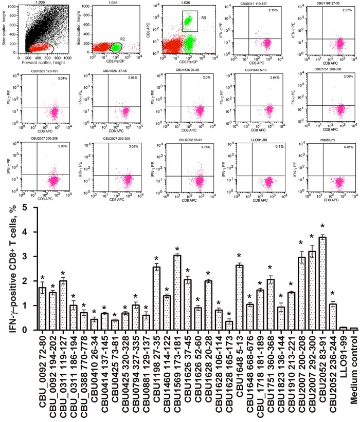
Figure 3.
Quantification of peptide-specific interferon γ (IFN-γ)–producing CD8+ T cells by intracellular cytokine staining and flow cytometry. Twenty micrograms of each peptide was used to stimulate 1 × 10⁶ lymphocytes obtained from 5 mice 10 days after challenge with Coxiella burnetii. After stimulation of lymphocytes for 24 hours with peptides, the expression of IFN-γ in CD8+ T cells was measured by flow cytometry. T cells cultured with Listeriolysin O peptide (LLO91–99) were used as a negative control. The data are representative of 3 independent experiments, and the average percentages of double-positive cells among the T cells are indicated in the top right corners. The mean values (±SDs) from the results of 3 independent experiments are also shown. *P < .05 for comparison of T cells stimulated with epitope peptides to T cells cultured with LLO91–99. Abbreviations: APC, allophycocyanin; PE, phycoerythrin; PerCP, peridinin chlorophyll protein.

