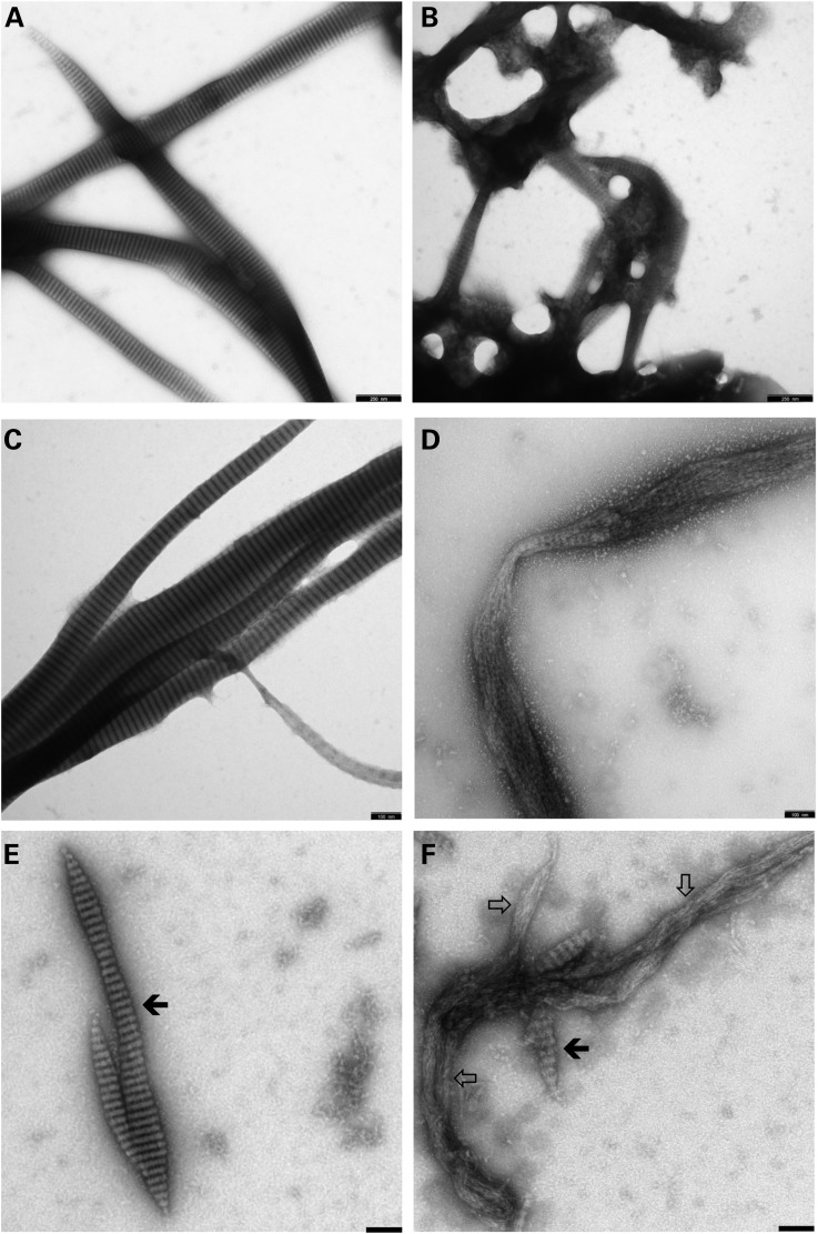
Figure 4.
LMNB2 protein assembly. Wild-type lamin B2 (A, C, E) and the mutant H157T-lamin B2 (B, D, F) were assembled in (A–D) 25 mm Mes-NaOH (pH 6.5), 150 mm NaCl, 25 mm CaCl2; and (E and F) 25 mm Tris–HCl (pH 8.5), 150 mm NaCl, 25 mm CaCl2, at room temperature for 45 min (A and B) and 60 min (C–F), respectively. The open arrows in (F) point to extended regions within the filamentous strands formed by the mutant protein that do not exhibit any ordered lateral organization; the filled arrows in (E) point to regions exhibiting the typical 24.5-nm repeat pattern (11) that on occasion is also seen within restricted regions in the mutant polymer (F). Refer to Results section for further explanation. Scale bars: (A and B) 250 nm, (C–F) 100 nm.

