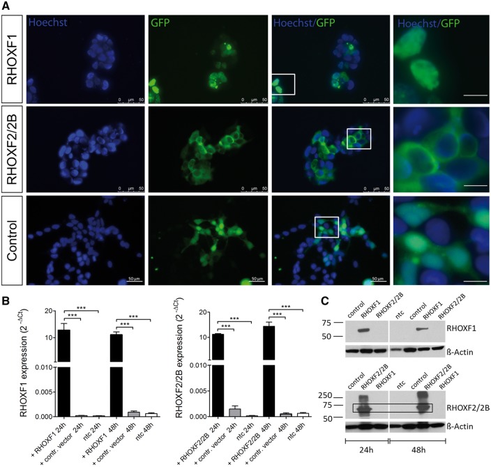
Figure 1.
RHOX expression in transiently transfected HEK293 cells. (A) Immunofluorescent imaging of HEK293 cells 48h after transfection. GFP signal reflects RHOX expression while nuclei were counterstained with Hoechst. Scale bar: 10 µm (right row). (B) RHOX expression after 24h and 48h of transfection (n = 8). Total RNA was isolated, reverse transcribed, and amplified with specific primers. Values were normalized to endogenous GAPDH. ***P < 0.001. (C) Western Blot analysis of RHOX after 24h and 48h of transfection.

