
Figure 4. Western blot analysis of UGTB10-over-expressing cell lines.
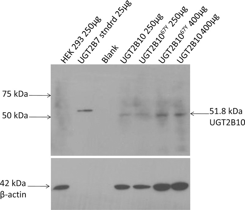
PVDF membrane was probed with 1:500 UGT2B antibody for 1 h at 23°C followed by donkey anti-goat IgG conjugated to horseradish peroxidase (1:4500) for 45 min at 23°C. Lane 1, HEK 293 (250 µg); lane 2, UGT2B7 protein standard (335 ng); lane 3, blank; lane 4, UGT2B10-over-expressing cell line (250 µg); lane 5, UGT2B1067Y-over-expressing cell line (250 µg); lane 6, UGT2B1067Y-over-expressing cell line (400 µg); lane 7, UGT2B10-over-expressing cell line (400 µg).