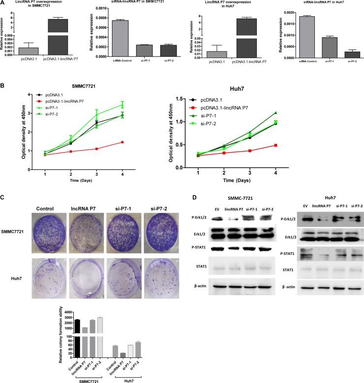
Figure 3. LincRNA P7 inhibited the proliferation of HCC cells in vitro.
(A) LincRNA P7 overexpression and knockdown in HCC cells lines. The expression level of lincRNA P7 was robustly enhanced or reduced when the cell lines were treated with pcDNA3.1-LincRNA P7 or with two siRNAs that target lincRNA P7, respectively. Empty vector and the siRNA control were used as the negative controls. (B) MTT assays were used to determine the cell viability of si-LincRNA P7- and pCDNA- LincRNA P7-transfected SMMC7721 and Huh7 cells. Cell number was determined every 24 h for a total of 96 h using CCK-8 assays. The results are shown as the means ± s.e.m. from three independent experiments. *P < 0.05, compared with the control by two-sided t-test. (C) Colony-forming assay was conducted to determine the effect of lincRNA P7 on the proliferation in SMMC7721 and Huh7 cells. (D) LincRNA P7 regulates the STAT1 and MAPK signaling pathway. Immunoblot analysis showed that the phosphorylation of STAT1 (pSTAT1) and Erk1/2 (pErk1/2) was significantly repressed in lincRNA P7-overexpressing SMMC7721 and Huh7 cells. In contrast, the phosphorylation of these proteins was rescued in si-lincRNA P7-transfected SMMC7721 and Huh7 cells.

