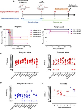
Fig. 1. Pregnant rats are more susceptible to death after RVFV infection, with virus homing to the liver and placenta.

(A) Experimental design for E14 SD rats infected with RVFV. After delivery at E22, dams and pups were not disturbed until 5 days postdelivery (13 dpi). Euthanasia of surviving dams and pups occurred 18 dpi (10 days postdelivery). (B) Survival of RVFV-infected pregnant dams and nonpregnant SD rats (n = 3 to 6 per dose). The shaded area represents the 2- to 6-day clinical window when lethally infected pregnant rats were euthanized owing to severe disease. (C) vRNA (qRT-PCR; left) and infectious virus (VPA; right) in tissues from pregnant rats that succumbed (red squares; n = 17) between 2 and 6 dpi. (D) vRNA in tissue samples from pregnant rats that survived infection (blue circles; n = 11) and were euthanized 18 dpi. Placenta samples (open blue circles) were obtained at day of delivery (8 dpi). (E) Infectious virus measured by VPA in placental samples obtained from lethally infected (red squares) and surviving (blue circles) rats at the indicated dpi. CLN, cervical lymph node. Dotted horizontal lines represent the limits of detection (LOD) of the qRT-PCR (0.1 PFU/ml eq) and VPA (50 PFU). ND, not detected (below the LOD); PFU, plaque-forming unit; PFU/ml eq, PFU per milliliter equivalents; VPA, viral plaque assay; vRNA, viral RNA.
