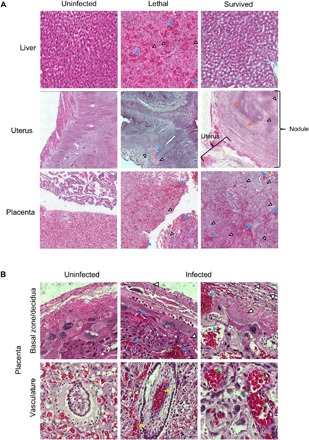
Fig. 2. RVFV causes pathology within the liver, uterus, and placenta of pregnant dams.

H&E staining within the indicated tissues. (A) Images (20×) of liver, uterus, and placenta. (B) Images (60×) of placenta. Blue, white, yellow, orange, and green arrowheads highlight evidence of hemorrhaging, necrosis, vascular/perivascular congestion, calcification, and nucleated red blood cells, respectively.
