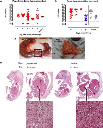
Fig. 3. Infection of pregnant dams results in direct transmission of RVFV to the peritoneum and brain of pups.

Pups delivered from (A) lethally infected and (B) surviving pregnant rats were tested for vRNA within the peritoneal cavity (left) and brain (right). In (A), the x axis represents the day the dams were euthanized owing to severe disease. In (B), the x axis represents the day after delivery, with day 10 representing surviving pups euthanized at the end of the study. For both graphs, red square data points indicate pup demise and blue circle data points indicate pup survival. Open data points are pup brain tissues; all closed data points are pup peritoneal cavity. (C) Photographic evidence of fetal resorption within the uterus of one of three dams that succumbed to RVFV infection. (D) Images (10×) of whole pups were examined for histological changes. H&E staining of a whole pup from a dam that succumbed to infection (right) or a corresponding uninfected control rat (left) euthanized at the same day of gestation. Blue, white, and black arrowheads highlight evidence of hemorrhaging, necrosis, and altered intestinal structure, respectively.
