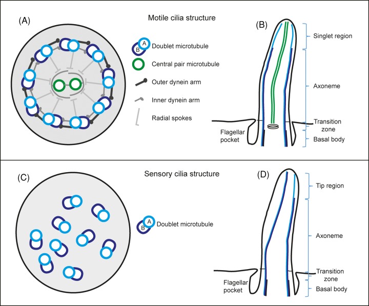
Figure 2. Schematic drawing of eukaryotic flagella ultrastructure.
(A) Cross-sectional view of a motile cilium seen from the proximal end containing the canonical nine doublet microtubules and two central pair microtubules. (B) A motile cilium in longitudinal view showing the different regions along its length. (C) A cross-sectional view of a sensory cilium showing the variable orientation of the nine doublet microtubules and lack of central pair microtubules. (D) A sensory cilium and its different regions along its length.

