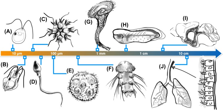
Figure 1. Unity and diversity of ciliary systems.
The same fundamental structure occurs in the tiniest of microorganisms as well as ciliated tissues, but exhibits drastic differences in number and localisation. Examples include (A) the algal biflagellate Chlamydomonas reinhardtii [4], (B) a quadriflagellate Prasinophyte alga Pyramimonas sp. [12], (C) rosette-forming choanoflagellates [13,14], (D) human sperm [15], (E) the spherical alga Volvox carteri [16,17], (F) the ciliated larvae of the marine annelid Platynereis dumerilii which have segmental multiciliated cells, and long, stiff chaetae [18], (G) the trumpet-shaped ciliate Stentor coeruleus [19], (H) ciliated epithelia of Xenopus laevis embryos [20], (I) ependymal cilia in mouse brain ventricles which direct cerebrospinal fluid flows [8] (cilia are localised to shaded region) and (J) multiciliated columnar cells in the human trachea [10,21].

