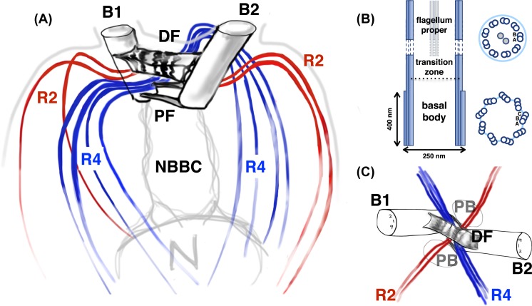
Figure 2. The Chlamydomonas flagellar apparatus.
(A) Schematic showing the cytoskeletal architecture of basal bodies (B1,2), microtubular roots (two-membered rootlets R2 and four-membered rootlets R4), and fibrous/contractile connections (NBBCs to the nucleus, proximal and distal striated fibres PF and DF). (B) Longitudinal section of a flagellum showing a structural change from triplet microtubules to doublets, two characteristic cross-sections are shown: one through the basal body and the second through flagellum proper. (C) Top view, highlighting radial symmetries in the flagellar apparatus, the locations of the two PB, the cruciate arrangement of microtubule bundles and the DF connecting specific microtubule doublets in the mature basal bodies (B1,2),

