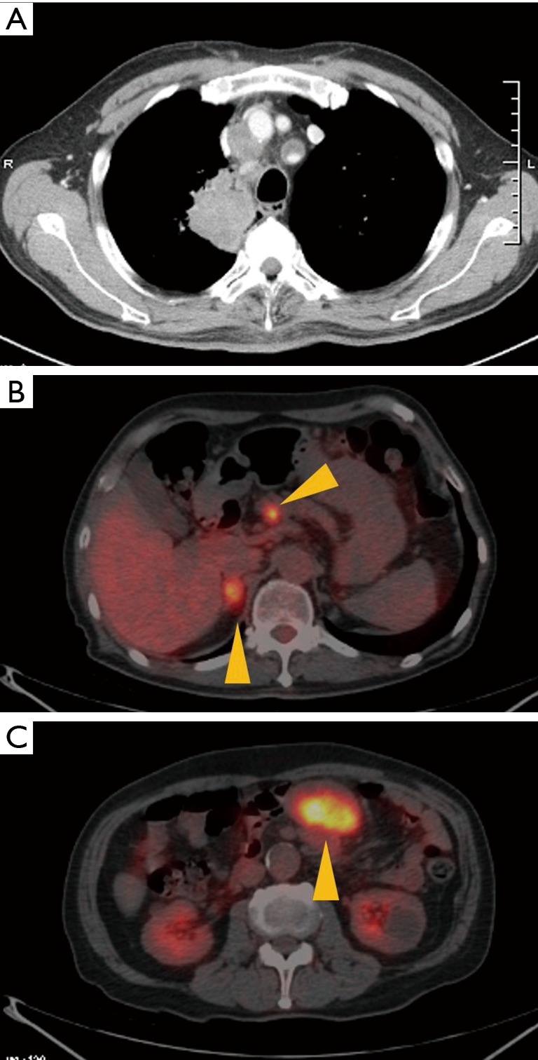
Figure 1.

Baseline chest computed tomography scan and PET scan of patient in case 1. (A) The diameter of the tumor in the right upper lung was approximately 56 mm, and mediastinal lymphadenopathy was observed. Accumulation of FDG on (B) the right adrenal gland, pancreas and (C) mesentery indicated the presence of metastasis. PET, positron emission tomography; FDG, fluorodeoxyglucose.
