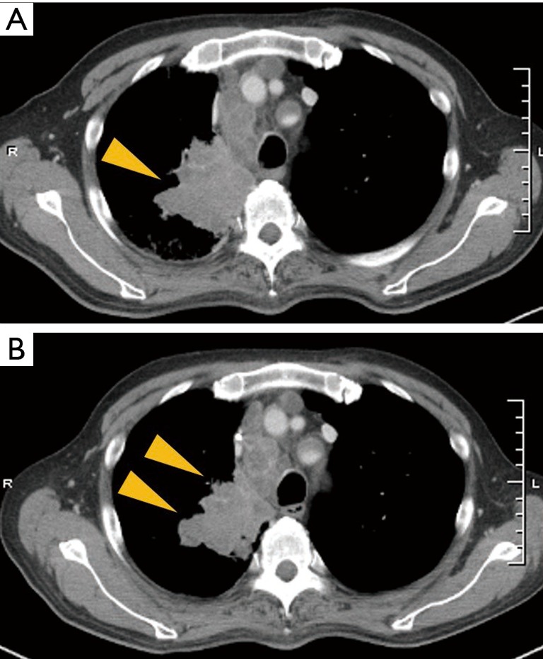
Figure 3.

Radiographical changes of the patient in case 1 during the pembrolizumab treatment (A) after treatment with erlotinib, CT scan showed a greater increase in the size of tumor in the right upper lobe and mediastinal lymphadenopathy, compared with the size shown in the initial CT scan; (B) twenty-one days after beginning pembrolizumab treatment, CT scan showed a reduced tumor size in the primary lesion of the right lung. CT, computed tomography.
