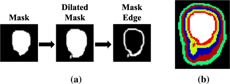
Fig. 2.

The schematic 2D representation of the technique used to extract the voxel values around the edges of the simulated bladder. a represents the procedure for extracting the voxel values, while b shows a 2D representation of all the dilated 3D regions around the bladder (i.e. the white region) from 2 voxels to 10 voxels with a step size of 2 voxels
