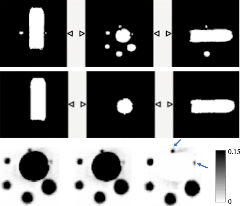
Fig. 9.

The NEMA bottle phantom used for the validation. The first row shows the MRAC image of the phantom and the middle row is the segmented bottle from the MRAC image, while the last row is showing the coronal view of the reconstructed images at three full iterations with a 4-mm Gaussian filter for OSEM, OSEM+PSF and OSEM+PSF+BC reconstructions, respectively. The blue arrows in the reconstructed images are pointing to the spheres in which there is visual improvement
