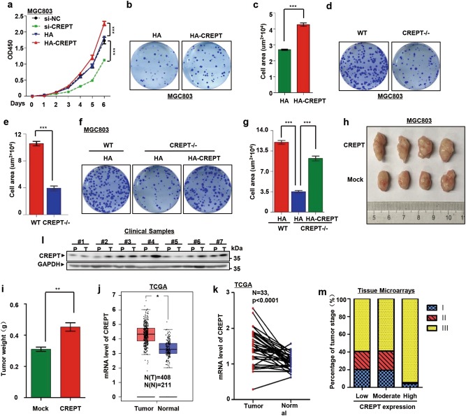
Fig. 1. CREPT promotes gastric cancer cell proliferation and is highly expressed in human gastric cancers.
a CREPT promotes the growth of MGC803 cells. Wild-type (WT), CREPT depletion (si-CREPT), an siRNA vector control (si-NC), CREPT overexpression (HA-CREPT), and vector control (HA) cells were used to measure the cell viability (presented as OD450 values). Results are represented as mean ± SD from three independent repeats. b, c Overexpression of CREPT promotes colony formation. Colonies formed by MGC803 cells stably overexpressing HA-CREPT were stained (b) with crystal violet and counted in three independent experiments (c). For each well, 500 cells were seeded and allowed for growth for 10 days. HA presents control cells with empty vector; ***p < 0.001. d, e Deletion of CREPT inhibits the colony formation. CREPT was deleted by a CRISPR/Cas9 system in MGC803 cells (CREPT–/–). For each well, 1000 cells were seeded and allowed for growth for 10 days. Colonies were stained (d) and quantitated from three independent experiments (e); ***p < 0.001. f, g Exogenous expression of CREPT rescues CREPT deletion effect on the colony formation. HA-CREPT was induced into MGC803 cells where endogenous CREPT was deleted by a CRISPR/Cas9 system. For each well, 1000 cells were seeded and colonies were stained (f). A quantitative presentation of colonies formation is shown (g) from 3 independent experiments; ***p < 0.001. h, i CREPT promotes the tumor growth. 1 × 10⁶ of mock and CREPT overexpressed MGC803 cells were injected into the flanks of NSG mice (n = 4). Mice were killed at day 21 and tumor weight was measured. Data are represented as mean ± SD. j CREPT is highly expressed in gastric cancers. Boxplots show the mRNA level of CREPT in tumor and normal tissues. Data were obtained from TCGA and GTEx database; *p < 0.05. k Elevated CREPT mRNA is showed in the paired gastric cancer and normal tissues. Levels of CREPT mRNA from paired tumor and normal tissues in the same patients were obtained from TCGA database. l The protein level of CREPT is increased in gastric cancers. A western blot was performed for gastric cancer samples from 7 patients. P refers to the paired non-tumor tissue and T refers to the tumor tissue from the same patient. GAPDH was used as a loading control. m A statistics of CREPT expression in different gastric cancer stages. Gastric cancers were grouped into three stages and the level of CREPT was categorized as low, moderate, and high from IHC results. Totally, 357 patients were observed

