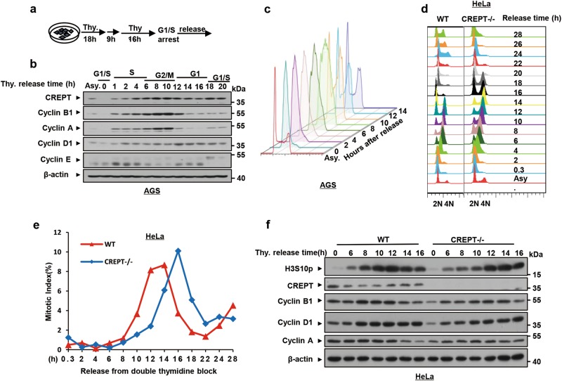
Fig. 2. The expression of CREPT correlates with the cell cycle progression.
a An experimental workflow shows the process of synchronization into G1/S phase. Thymidine (Thy.) was added for 18 h before a 9 h release and was re-added for another 16 h. This double thymidine block (DTB) protocol allows cell synchronized at the G1/S phase. b The expression of CREPT alters along with cell cycle stages. The protein level of CREPT was examined by western blots from cells in different cell cycle stages. Asynchronous (Asy.) and synchronized cells were used. Other cell cycle-related proteins were used as markers to indicate the cell cycle stage. β-Actin was used as a loading control. c FACS analyses show the cell cycle in different stages after release from DTB. A total number of 2 × 10⁴ cells were used for FACS analyses. d CREPT deletion extends the cell cycle. Synchronized wild-type (WT) and CREPT deletion (CREPT–/–) HeLa cells were released and collected at the indicated time points. Cells were stained with PI for FACS analyses. e Deletion of CREPT leads to a cell cycle delay of 2 h. A phosphorylation antibody against H3S10p and Alexa Fluor® 488 conjugate secondary antibody were used to analyze the positive cell population by FACS. The mitotic index was presented by the percentage of H3S10p-positive cells. f Deletion of CREPT postpones the phosphorylation of H3S10p. A western blot was performed to demonstrate the level of H3S10p for the WT and CREPT–/– cells released from synchronization. Other cell cycle-related proteins were examined to demonstrate the cell cycle correlation

