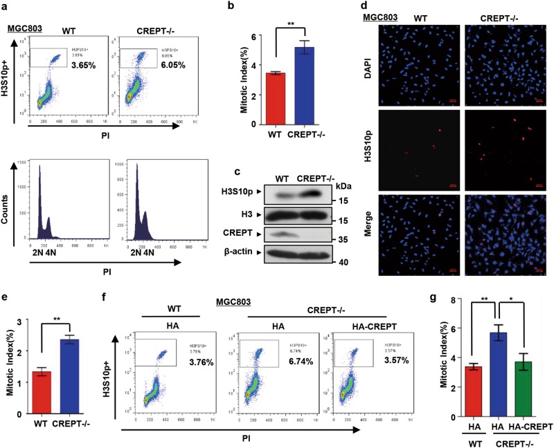
Fig. 3. Deletion of CREPT leads to significant mitotic cell accumulation.
a–c The mitotic index increases in CREPT deletion cells. Cells were harvested and stained with PI and H3S10p antibody. FACS analyses showed increased percentage of H3Ser10-positive cells when CREPT was deleted (CREPT–/–) (a). A quantitative presentation of the mitotic index from three independent experiments (b) **p < 0.01. A western blot showed the level of H3Ser10 phosphorylation. H3 and β-actin were used as loading controls (c). d Immunofluorescence visualization of mitotic cells. Wild-type (WT) and CREPT deletion (CREPT–/–) MGC803 cells were stained for H3S10p (red) and DAPI (blue). Scale bars, 100 μm. e The percentage of H3S10p-positive cells. Approximately 4000 cells were counted per sample in three independent experiments; **p < 0.01. f Overexpression of HA-CREPT rescues the CREPT deletion-induced mitotic arrest. HA-CREPT was stably induced into CREPT deletion cells. g A quantitative presentation of the mitotic index. Three independent experiments were analyzed by Student's t-test; **p < 0.01, *p < 0.05

