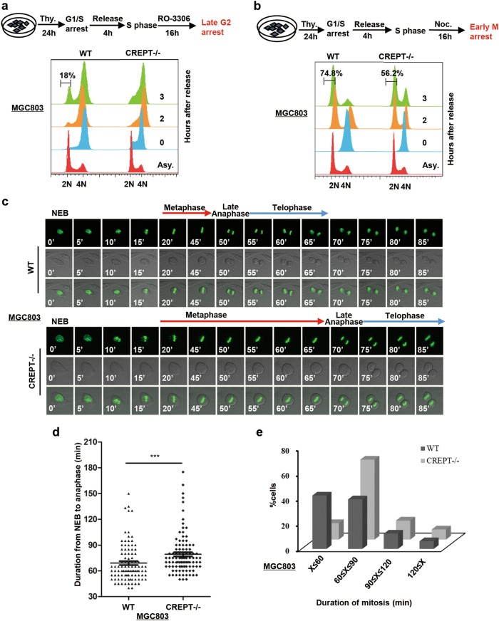
Fig. 4. Deletion of CREPT prolongs mitotic progression in gastric cancer cells.
a An experimental workflow shows the process of synchronization cells into late G2 phase by thymidine (Thy.) and CDK1 inhibitor (RO-3306) (Top). Deletion of CREPT induces cell to arrest at the G2/M phase. Wild-type (WT) and CREPT deletion (CREPT–/–) MGC803 cells were treated by thymidine and RO-3306 and released into normal fresh medium. DNA contents were analyzed by FACS (2 × 10⁴ cells/sample) (bottom). b An experimental workflow shows the process of synchronization cells into the early M phase by thymidine (Thy.) and nocodazole (Noc.) (Top). Deletion of CREPT delays the cell entry into the next G1 phase. Thymidine- and nocodazole-treated cells were released and analyzed for DNA content by FACS (2 × 10⁴ cells/sample) (bottom). c CREPT deletion cells exhibit prolonged mitosis. Selected frames from a time-lapse microscopy for GFP-H2B infected wild-type (WT) and CREPT deletion (CREPT–/–) MGC803 cells are shown. NEB nuclear envelope breakdown. d A dot pot shows the duration from NEB to telophase in MGC803/GFP-H2B cells where CREPT was deleted (CREPT–/–). Wild-type (WT) cells were used as a control; n = 99. e Distribution of cells during different mitotic stages. About 60% of CREPT deletion cells need about 80 min to finish mitosis, while most of the wild-type cells need less than 60 min. The duration of the mitotic time was categorized into four groups; n = 99

