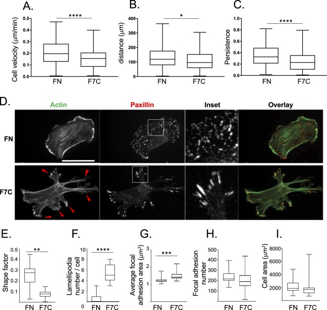
Figure 5.
Fbln7-C affects focal adhesion area and actin filaments to inhibit cell motility. (A–C) Cell motility on fibronectin or Fbln7-C-coated dishes stimulated with VEGF and analyzed using time-lapse imaging. Cell motility is evaluated by (A) cell velocity, (B) distance, (C) and persistence. N = 3 n > 150, ****P < 0.0001, **P < 0.01, *P < 0.01. (D–I) Cell morphology differences between cells on fibronectin and Fbln7-C-coated dishes evaluated by staining for focal adhesion sites (paxillin; red) and actin filaments (phalloidin; green). (D) Immunofluorescence staining. Cell shape is evaluated by (E) shape factor and (F) number of lamellipodia. Focal adhesion area is evaluated by (G) average focal adhesion area, (H) focal adhesion number and (I) cell area. FN: Fibronectin, F7C:Fibulin7-C, scale bar: 100 μm, N = 3 n > 80, ****P < 0.0001, **P < 0.01, *P < 0.01.

