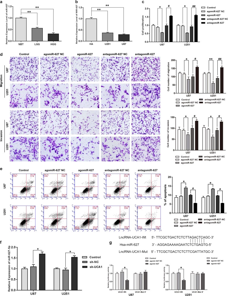
Fig. 2. MiR-627-5p acted as a tumor suppressor in glioma cells and was down-regulated by UCA1.
a MiR-627-5p expression in normal brain tissues (NBTs), Grade I–II glioma tissues, Grade III–IV glioma tissues. Error bars represent as the mean ± SD (n = 5, each group). **P < 0.01, ##P < 0.01. b MiR-627-5p expression in human normal astrocytes and GBM cell lines (U87 and U251). Error bars represent as the mean ± SD (n = 5, each group). **P < 0.01, ##P < 0.01. c Effect of miR-627-5p on cell proliferation of U87 and U251 cells. d Effect of miR-627-5p on cell migration and invasion of U87 and U251 cells. e Effect of miR-627-5p on cell apoptosis of U87 and U251 cells. Error bars represent as the mean ± SD (n = 3, each group). *P < 0.05, #P < 0.05. Scale bars represent 20 μm. f Relative expression of miR-627-5p after cells transfected with the expression of UCA1 changed. Error bars represent as the mean ± SD (n = 3, each group). *P < 0.05. g Relative luciferase activity was performed by dual-luciferase reporter assay. Error bars represent as the mean ± SD (n = 3, each group). *P < 0.05. Scalebars represent 20 μm

