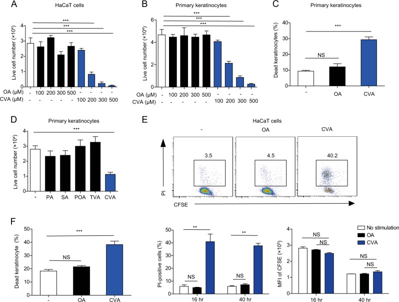
Fig. 5. CVA induces keratinocyte death in the skin.
a, b Number of live cells in cultures of HaCaT cells (a) and primary keratinocytes isolated from the skin of new born mice (b) 16 h after stimulation with indicated concentration of OA or CVA was calculated using trypan blue exclusion test (n = 3 per group). c Primary keratinocytes were stimulated for 6 h with 300 μM of OA or CVA, stained with propidium iodide (PI) and analyzed by flow cytometry for the proportion of PI-positive (i.e., dead) cells (n = 4 or 5 per group). d Number of live cells in primary keratinocytes 16 h after stimulation with 300 μM of PA, SA, POA, TVA, or CVA was calculated using trypan blue exclusion test (n = 3 per group). e HaCaT cells were labeled with CFSE and stimulated or not with 300 μM of OA or CVA for 16 or 40 h. Cells were then stained with PI and analyzed by flow cytometry. f Epidermal cells of mice that received topical application of ethanol (control) or 45 mM of OA or CVA to the dorsal skin for 6 h were stained with anti-CD45.2, CD49f, and PI and analyzed by flow cytometry for PI-positive (i.e., dead) CD45.2−CD49f+ keratinocytes. Error bars indicate SD. **P < 0.01, ***P < 0.001. NS not significant, OA, oleic acid, CVA cis-vaccenic acid, PA palmitate, SA stearate, POA palmitoleate, TVA trans-vaccenic acid. Data are representative of more than two independent experiments

