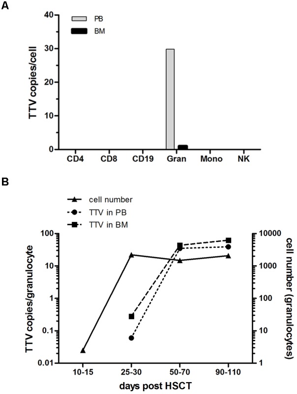
FIGURE 3.

TTV detection in leukocyte subsets and virus replication kinetics after transplantation. Leukocyte subsets including CD4+, CD8+, CD19+, CD15+ (granulocytes: Gran), CD33+ (monocytes: Mono) and CD56+ (natural killer cells: NK) were isolated by flow sorting from peripheral blood (PB) and bone marrow (BM) of 19 pediatric HSCT recipients. Individual cell subsets were tested for the presence and quantity of virus copies by a TTV-specific RQ-PCR assay. (A) The median of the peak TTV copy numbers per cell determined in the patients investigated is shown for the different leukocytes subsets. (B) The TTV replication kinetics in granulocytes isolated from PB and BM of the 19 patients investigated is shown by the median virus copy numbers per cell at the indicated time points post-HSCT. The y-axis on the right side indicates the granulocyte numbers per μl PB.
