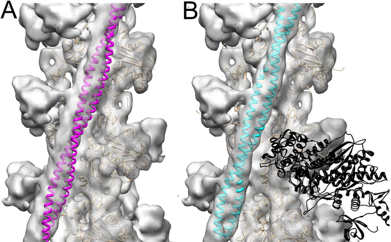
Figure 2.
C1 binding to the Tm cable traps it in the myosin rather than Ca2+-induced c-open structural state. (A) Atomic model of the cardiac open (c-open) structural state of the cardiac TF (Protein Data Bank ID code 5NOJ) (Risi et al., 2017) was rigidly docked into the 3D reconstruction of the C1-S structural class to show that Tm (magenta ribbons) does not fit into the portion of the electron density map that corresponds to the Tm cable. (B) The model of the myosin-Tpm-F-actin complex (von der Ecken et al., 2016) (Protein Data Bank ID code 5JLH) docked into the 3D reconstruction of the C1-S structural class without any perturbations unambiguously shows that Tm (cyan ribbons) position in the 3D reconstruction of the C1-S class corresponds to the myosin state of the TF. 3D reconstructions are shown as grey transparent surfaces. Actin molecules are shown as tan ribbons, while rigor bound myosin-S1 head is shown as black ribbons.

