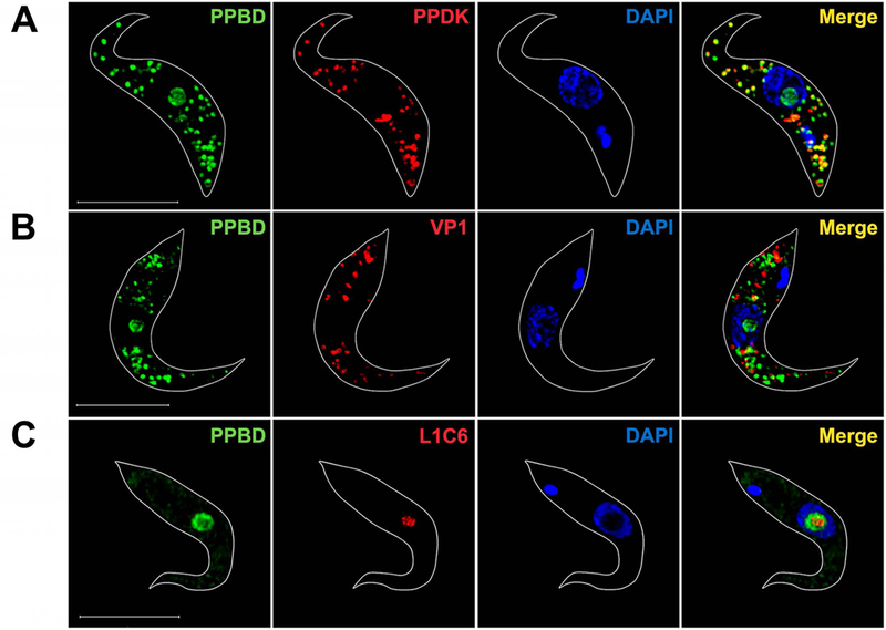
Fig. 1.

Super-resolution images of PPBD-labeled T. brucei PCF.
(A) PPBD (green) (8 µg/ml) localizes in the nucleolus, which is identified as the nuclear region not stained with DAPI (blue) and co-localizes (Merge, yellow) with antibodies against the pyruvate phosphate dikinase (PPDK, red) in the glycosomes.
(B) PPBD does not co-localize with antibodies against TbVP1 (red), the acidocalcisome marker.
(C) PPBD nucleolar localization coincides but does not superimpose to the labeling by nucleolar antibody L1C6 (red). The concentration of PPBD used in (C) was lower (2 µg/ml) to show only nucleolar labeling. Scale bars = 5 µm.
