Fig. 3.

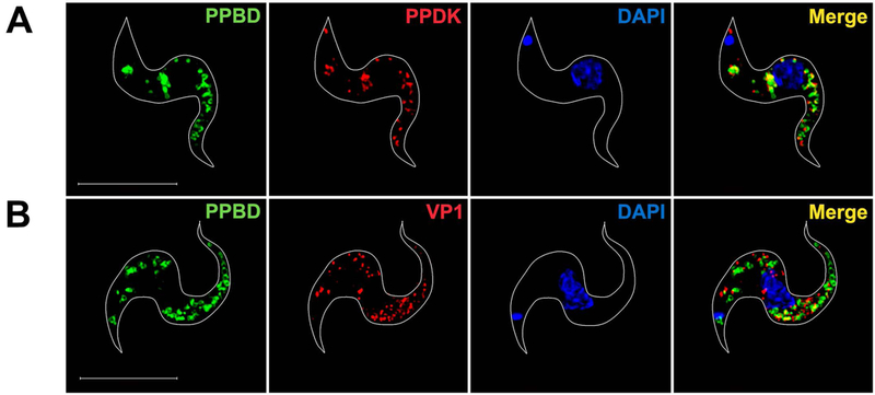
Super-resolution images of PPBD-labeled T. brucei BSF.
(A) PPBD (green) does not label the nucleolus and partially co-localizes (Merge, yellow) with antibodies against PPDK (red).
(B) PPBD does not co-localize with antibodies against TbVP1 (red). DAPI staining is in blue. Scale bars = 5 µm.
