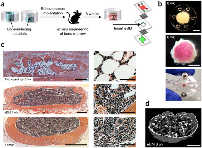
Figure 5.

Bone marrow-on-a-chip. (A) Workflow to generate a bone marrow-on-a-chip system. (B) Top, PDMS device containing bone-inducing materials before implantation. Center, white cylindrical bone with pink marrow within eBM 8 weeks after implantation. Bottom, in vitro bone marrow (BM) chip microdevice. (C) H&E-stained sections of the eBM formed in the PDMS device with two openings (top), or one lower opening (center) at 8 weeks following implantation, bone BM in a normal adult mouse femur (bottom). (D) 3-D reconstruction of micro-CT data from eBM 8 weeks after implantation (from Ref. [263] with permission).
