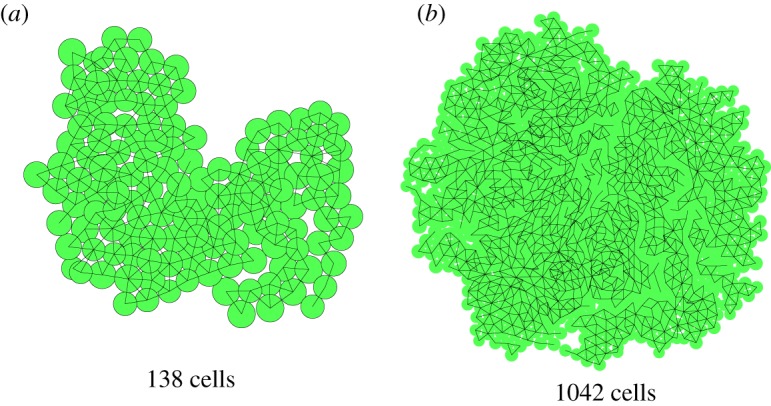
Figure 8.

The same sample after 175 (a) and 250 steps (b), both grown from a single cell. These images are taken from a single run, continued from the previous image. In (b), the perimeter of the circle representing the cell is left out, making the network of the cells stand out more clearly. The network has many branches and peninsula-like structures, giving it a fractal-like appearance.
