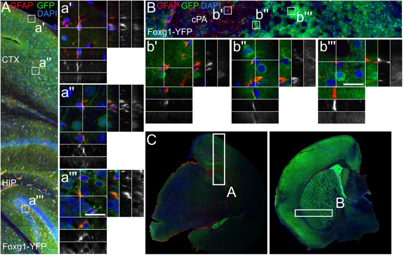
FIGURE 6.
GFAP-positive astrocytes partially derive from FOXG1-cre expressing progenitors. Lineage tracing of FOXG1-expressing cells with a YFP reporter mouse line demonstrates that GFAP expressing astrocytes derive from FOXG1-expressing progenitors in (A) cerebral cortex (CTX) and hippocampus (HIP) and in (B) the caudate putamen (cPA). Magnifications show single GFAP- and YFP-positive (GFAP+/YFP+) cells in (a’,a”) cerebral cortex, (a”’) hippocampus and (b”) caudate putamen. In (b’,b”’) GFAP+/YFP-negative cells are illustrated in the caudate putamen. Scale bar: 10 μm, n = 3. (C) Overview images of representative rostral and caudal forebrain section after immunofluorescence for GFAP and GFP show the region analyzed in (A,B) as indicated.

