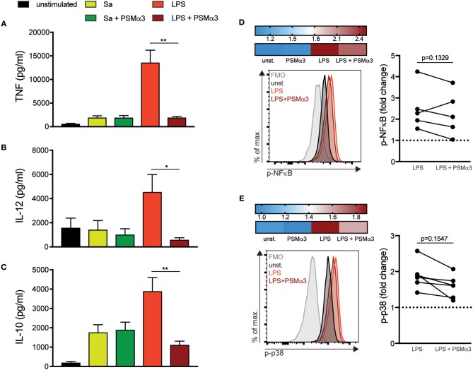
Figure 2.
PSMs impair pro- and anti-inflammatory cytokine secretion by moDCs. (A–C) MoDCs were treated with Sa lysate or LPS with or without PSMα3 for 6 h (A) or 24 h (B,C). The cell culture supernatants were collected and analyzed for TNF (A), IL-12 (B) and IL-10 (C) production by sandwich ELISA. The graphs show n ≥ 10 independent experiments (mean ± SD/SEM) performed in triplicates. (D,E) MoDCs were treated with LPS with or without PSMα3 or PSMα3 alone for 60 min. Thereafter, the cells were stained extracellularly with anti-CD11c and anti-HLA-DR antibodies, followed by fixation, permeabilization and subsequent intracellular staining using anti-phospho-NF-κB (D) and anti-phospho-p38 antibodies (E). Representative histogram overlays of p-NF-κB (D) and p-p38 (E) in DCs (gated on CD11c+HLA-DR+ cells). The heat map shows fold-change of p-NF-κB (D) and p-p38 (E) normalized to untreated DCs (unst.). The graphs show the statistical analysis of p-NF-κB (D) and p-p38 (E) from n = 5 different donors. *p < 0.05 or **p < 0.005, Kruskal-Wallis with Dunn's posttest or one-way ANOVA with Turkey's posttest.

