Figure 3.
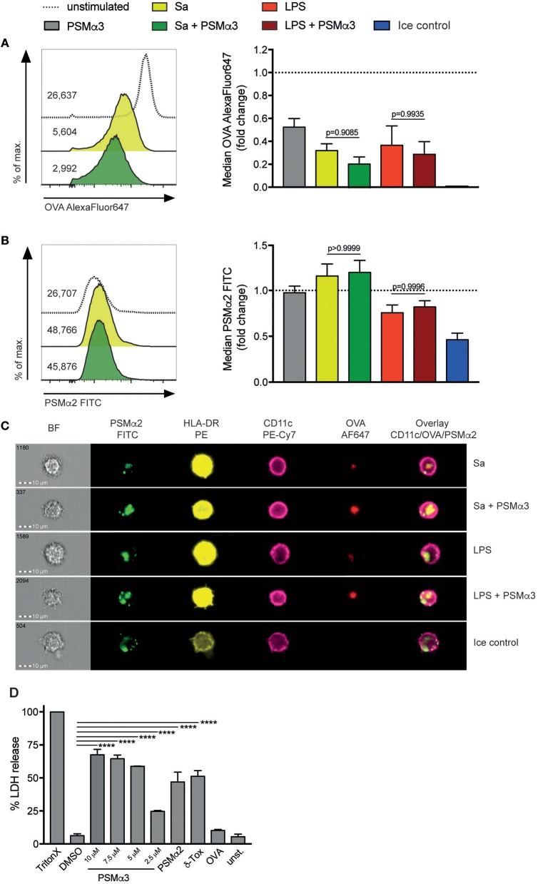
PSMs inhibit antigen uptake by moDCs and penetrate the membrane via transient pore formation. (A–C) MoDCs treated for 24 h with Sa lysate or LPS with or without PSMα3 or PSMα3 alone were incubated with AlexaFluor647-labeled OVA and FITC-labeled PSMα2 at 37°C or on ice for 30 min. (A,B) The uptake of OVA and PSMα2 by living CD11c+HLA-DR+ moDCs was assessed by flow cytometry. The histogram overlays (left) and the bar graphs (right, collected data) show the mean fluorescence intensity of OVA-AlexaFluor647 (A) or PSMα2-FITC (B), in case of the collected data as fold change of unstimulated cells (n ≥ 5, performed in triplicates, mean ± SEM). (C) The localization of OVA-AlexaFluor647 and PSMα2-FITC in CD11c+HLA-DR+ moDCs was analyzed by multispectral imaging flow cytometry (one representative experiment of n = 3 independent experiments; mean ± SEM). Representative bright field (BF) and fluorescence images of moDCs are shown for the different treatments. (D) MoDCs were either not treated (unst.) or incubated with 1% Triton X-100, 2% DMSO, PSMα3 (2.5 μM, 5 μM, 7.5 μM or 10 μM), 10 μM PSMα2, 10 μM δ-Toxin or 5 μg/mL OVA for 10 min. The LDH release was determined in the cell culture supernatants (one representative experiment of n = 3 independent experiments; mean ± SEM). ****p < 0.0001, one-way ANOVA with Turkey's posttest or Kruskal-Wallis with Dunn's posttest.

