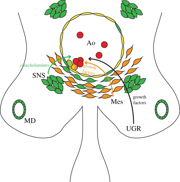
Figure 3.

Gata3 involvement in AGM haematopoiesis. Schematic diagram of a transverse section through an E11.5 AGM region, highlighting the cell compartments that express Gata3. Gata3-positive cells (green) are found within the endothelial layer (yellow) of the dorsal aorta (Ao), in the mesonephric duct (MD), within the subaortic mesenchyme (orange; Mes) and in the sympathetic nervous system (SNS). Blood cells are shown in red. The light green cell depicts the putative involvement of Gata3 in the endothelial-to-haematopoietic transition. Curly arrows illustrate contributions made by the different components of the microenvironment to EHT/HSC support, of which only catecholamines are currently known to be Gata3-dependent. UGR, urogenital ridges.
