
Figure 6.
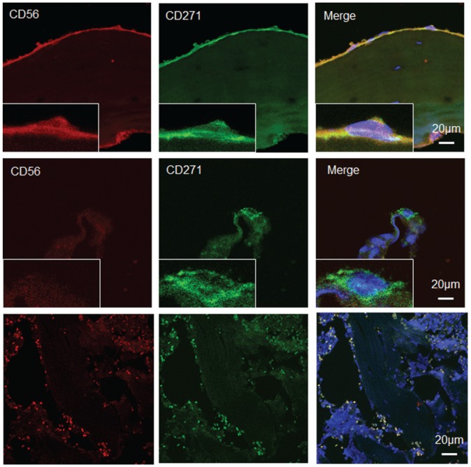
Co-localization of CD271 and CD56 in femoral head bone marrow cells. Double immunofluorescence staining with (top row) CD271 and CD56 identified CD271+CD56+ cells in the bone-lining region and (middle row) CD271+CD56– cells in the perivascular region. (Bottom row) In the negative control panel, very small cells have no DAPI staining (because of the absence of nuclei) but are autofluorescent in the red and green channels corresponding to cells, including red blood cells.