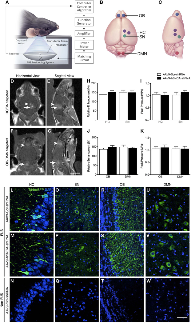
Fig. 1: MRI-guided FUS-induced BBB opening and transgene expression following FUS-mediated delivery of AAV9-hSNCA-shRNA and AAV9-Scr-shRNA to targeted brain regions.
(A) Schematic of the FUS experimental setup. (B,C) Selected focal spots in the HC, SN, OB and DMN (indicated by purple, green, blue and red dots, respectively) for FUS-mediated BBB disruption. (D,E) Following FUS, T1-weighted MR images were taken to confirm increased BBB permeability at selected focal spots in the HC and SN in one cohort of animals, and (F,G) in the OB and DMN in a second cohort. There was no significant difference in (H,J) enhancement level and (I,K) mean peak pressure required to induce BBB opening between AAV9-hSNCA-shRNA (black bar) and AAV9-Scr-shRNA treated animals (white bar) for both targeting schemes. (L-W) Representative confocal images (60X magnification) of brain sections prepared from AAV9-hSNCA-shRNA and AAV9-Scr-shRNA injected animals to detect turboGFP immunofluorescence. For both experimental groups, viral-mediated turboGFP was restricted to the FUS-treated side in the (L,M) HC and (O,P) SN, and bilateral (R,S) OB and (U,V) DMN. In contrast, turboGFP was not detectable on the contralateral hemisphere in the (N) HC and (Q) SN, nor in the untreated (T) OB and (W) DMN. Statistics: Paired, two-tailed, student’s t-test. Data represent the mean ± SEM. n = 4 per group. TurboGFP, green; DAPI, blue. Scale bars: (D-G) 5 mm; and (L-W) 20 μm.

