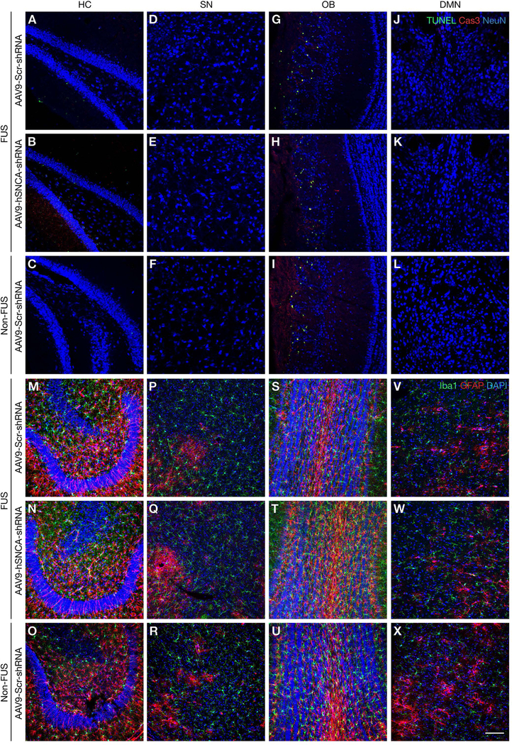
Fig. 5: MRI-guided FUS gene delivery does not induce neuronal apoptosis or inflammation.
(A-L) Representative confocal images of brain sections immunolabeled for TUNEL (green), cleaved caspase-3 (Cas3,red) and NeuN (blue). TUNEL- and cleaved caspase-3 positive cells were not detected in sonicated AAV9-Scr-shRNA or AAV9-hSNCA-shRNA-transduced areas of the (A, B) HC, (D, E) SN, (G, H) OB (relative to basal expression levels), and (J, K) DMN. (C, F, I, L) Immunostaining in non-FUS areas showed a similar pattern of apoptotic labeling. (M-X) Confocal images of brain sections depicting Iba-1(green), GFAP (red), and nuclei (DAPI, blue) immunolabeled cells. There was no difference in expression between the sonicated AAV9-hSNCA-shRNA and AAV9-Scr-shRNA treatment in the (M, N) HC, (P, Q) SN, (S, T) OB, and (V, W) DMN, as compared to immunolabeling in corresponding non-FUS regions (O, R, U, X). Scale bar: 100μm.

