Fig. 1.
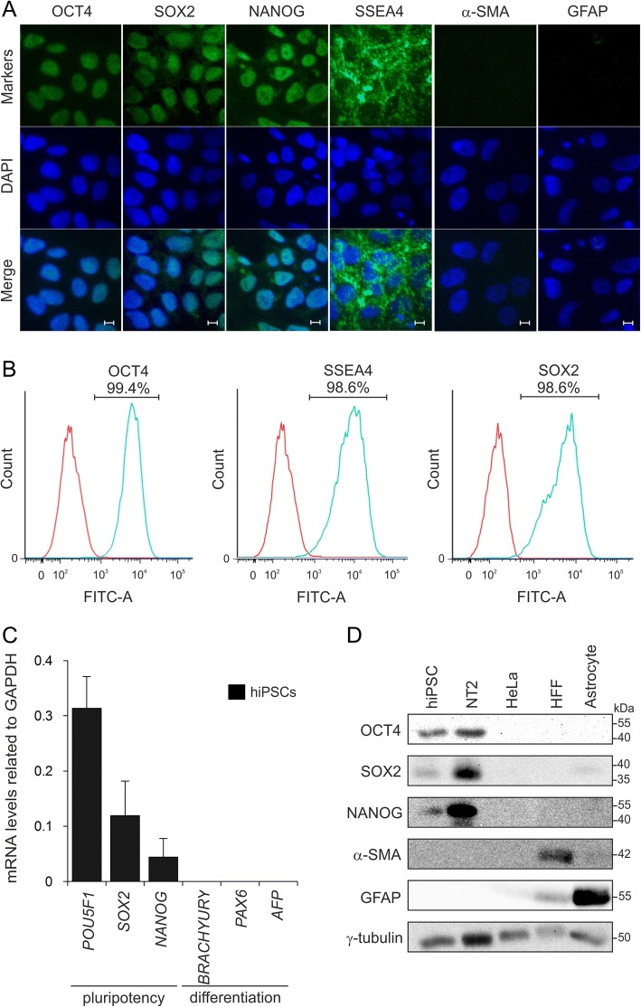
Long-term maintenance of undifferentiated hiPSCs. a Confocal imaging showed the expression of pluripotency markers (OCT4, SOX2, NANOG and SSEA4) and the absence of differentiation markers GFAP and α-SMA as ectodermal and mesodermal markers, respectively. Cell nuclei were stained with DAPI (blue). Scale bars, 10 μm. b Flow cytometry confirmed expression of OCT4, SSEA4 and SOX2 in hiPSCs with more than 98% of positive cells. c qPCR analysis for undifferentiated stem cell markers (POU5F1, SOX2 and NANOG) and early commitment to differentiation markers (BRACHYURY, PAX6 and AFP). GAPDH was used as an internal control. d Immunoblot analysis showing the specificity of antibodies and expression of markers. HeLa cells were used as negative control, HFF and astrocytes were used as positive control for α-SMA and GFAP, respectively

